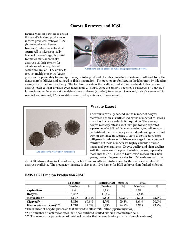
Embryo Morphology & Grading
Morphology roughly means “structure,” so embryo morphology describes the structure of the embryo as it develops.
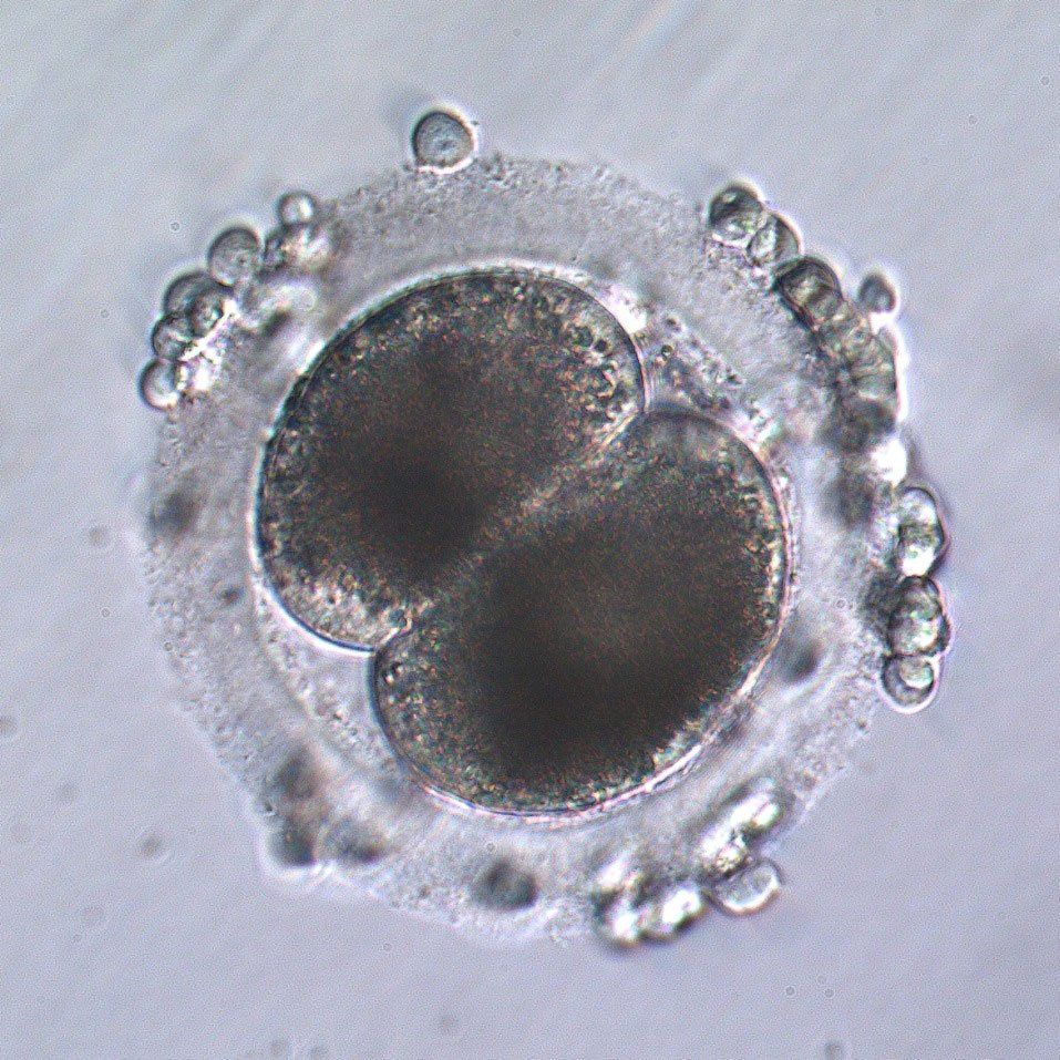
Early Embryo
Fertilization occurs in the mare’s oviduct and the first cell division takes place about 24 hours later. The cell cycle is approximately 24 hours and cell numbers double with each cycle.
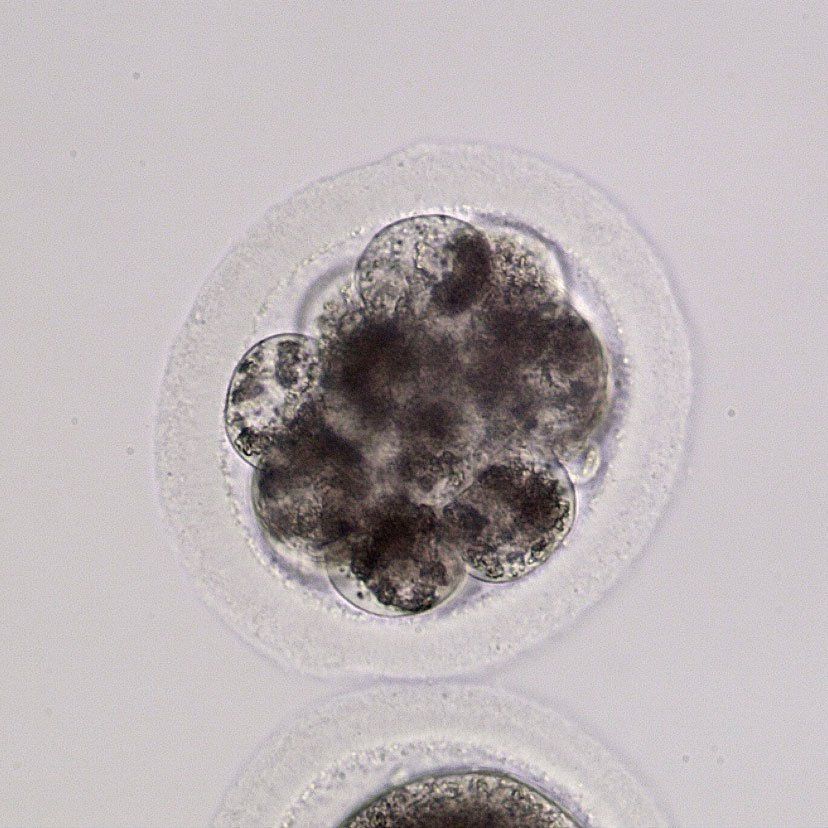
Morula
The equine embryo enters the uterus within about five and a half days after conception in the form of a morula. A morula is essentially a ball of cells surrounded by a clear structure called the zona pellucida. With each cell division in the oviduct, the cells divide without growing in size, so the embryo is still roughly the size of an oocyte at 160 microns (0.16mm or 7/1,000 of an inch) when it enters the uterus. At that time, the cells of the morula start drawing together, creating the compact morula.
Morula, 160 microns.
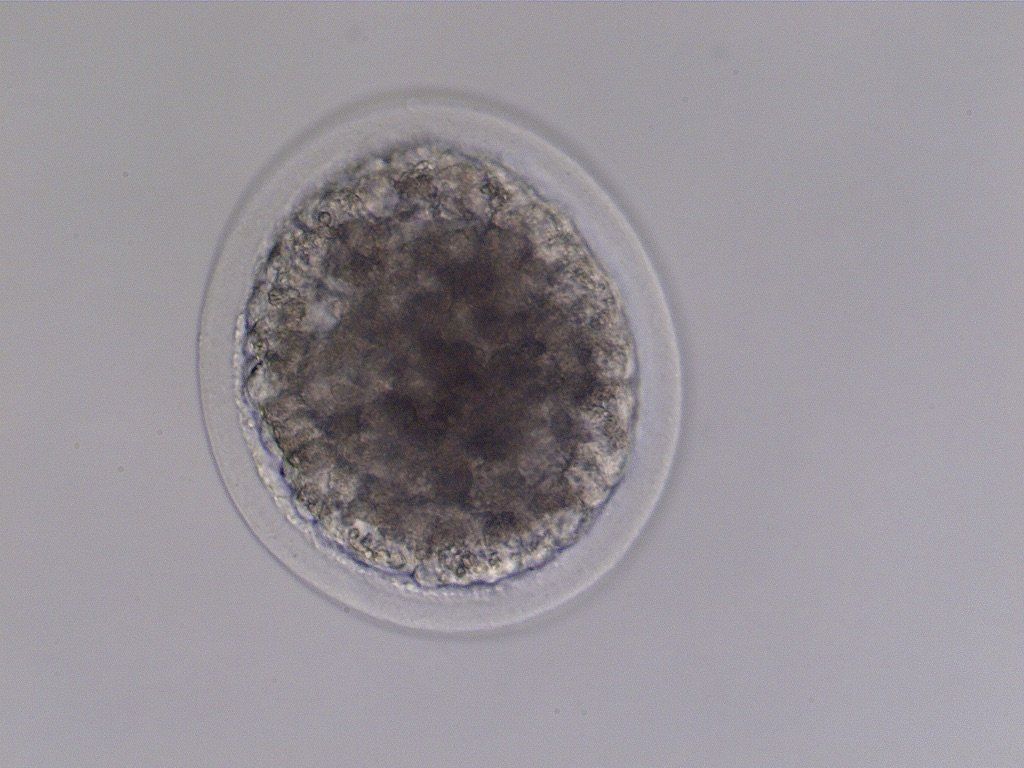
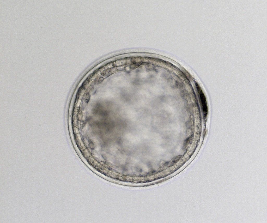
Early Blastocyst
At six to seven days post-fertilization, the compact morula starts organizing to create a layer of cells under the zona pellucida, called trophoblasts. As this layer forms, a fluid-filled cavity called the blastocoele begins forming in the center. The inner cell mass becomes apparent; this will later form the embryo proper, the future fetus. The embryo capsule forms between the zona pellucida and the trophoblast cell layer in the early blastocyst. The embryo now starts increasing in size causing the zona pellucida to thin
Early blastocyst, 210 microns
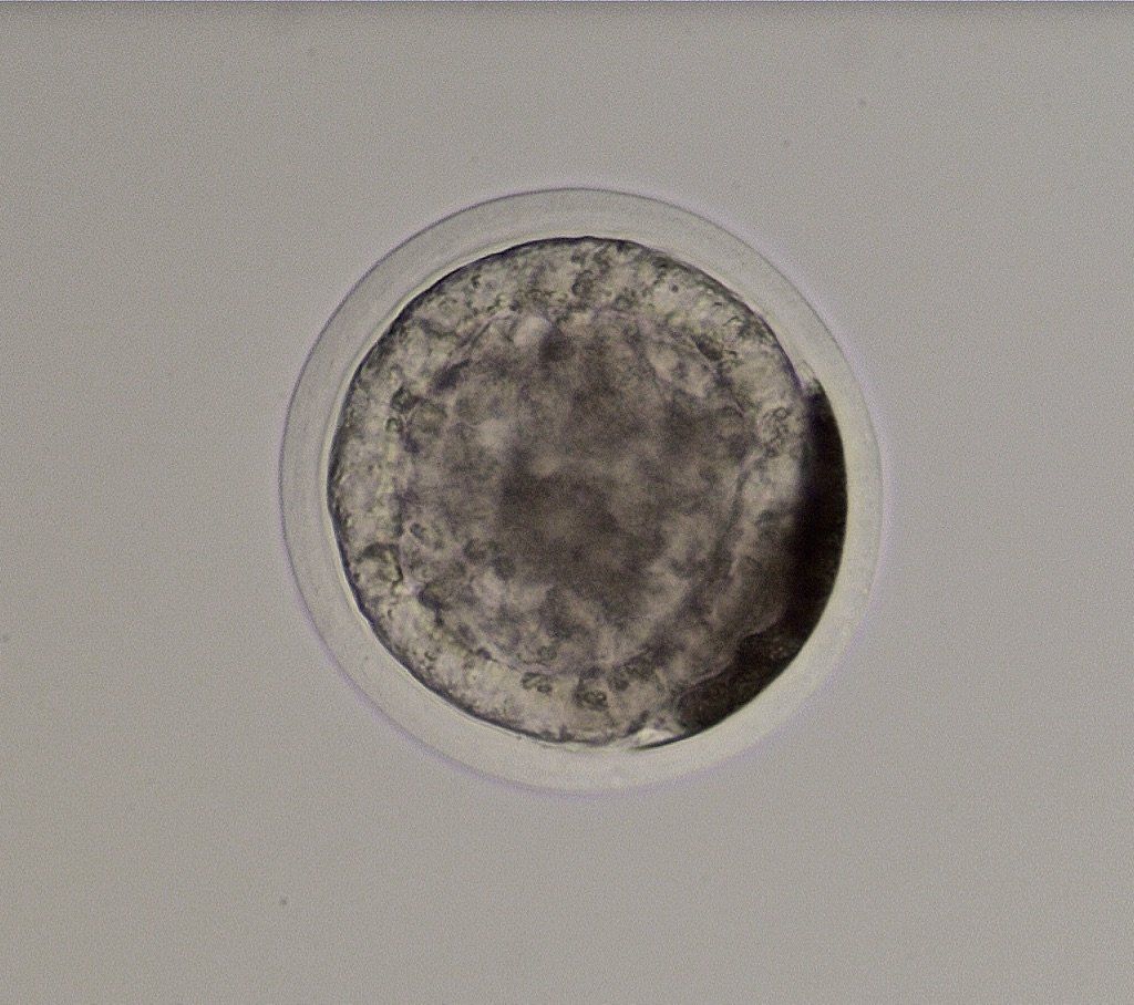
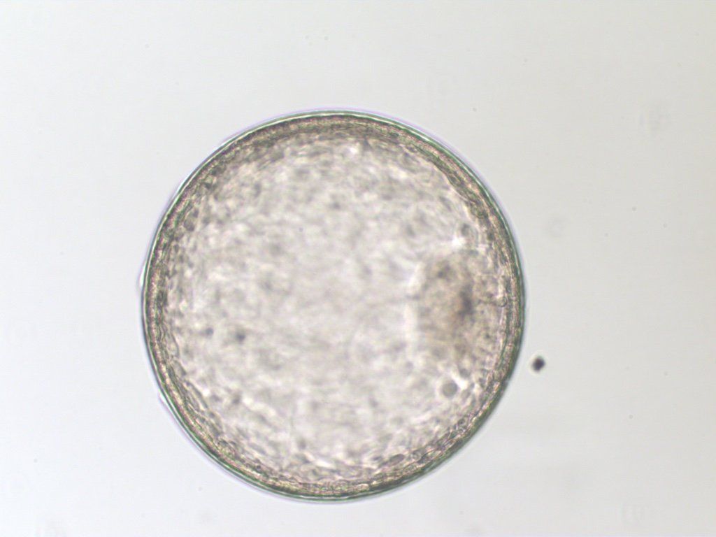
Expanded Blastocyst
Around day seven, the embryo starts growing rapidly in diameter as the blastocoele expands. The zona pellucida thins and flakes away. The inner cell mass becomes more prominent and extruded blastomeres that were trapped beneath the zona pellucida and on top of the capsule are shed.
Expanded blastocyst, 300 microns
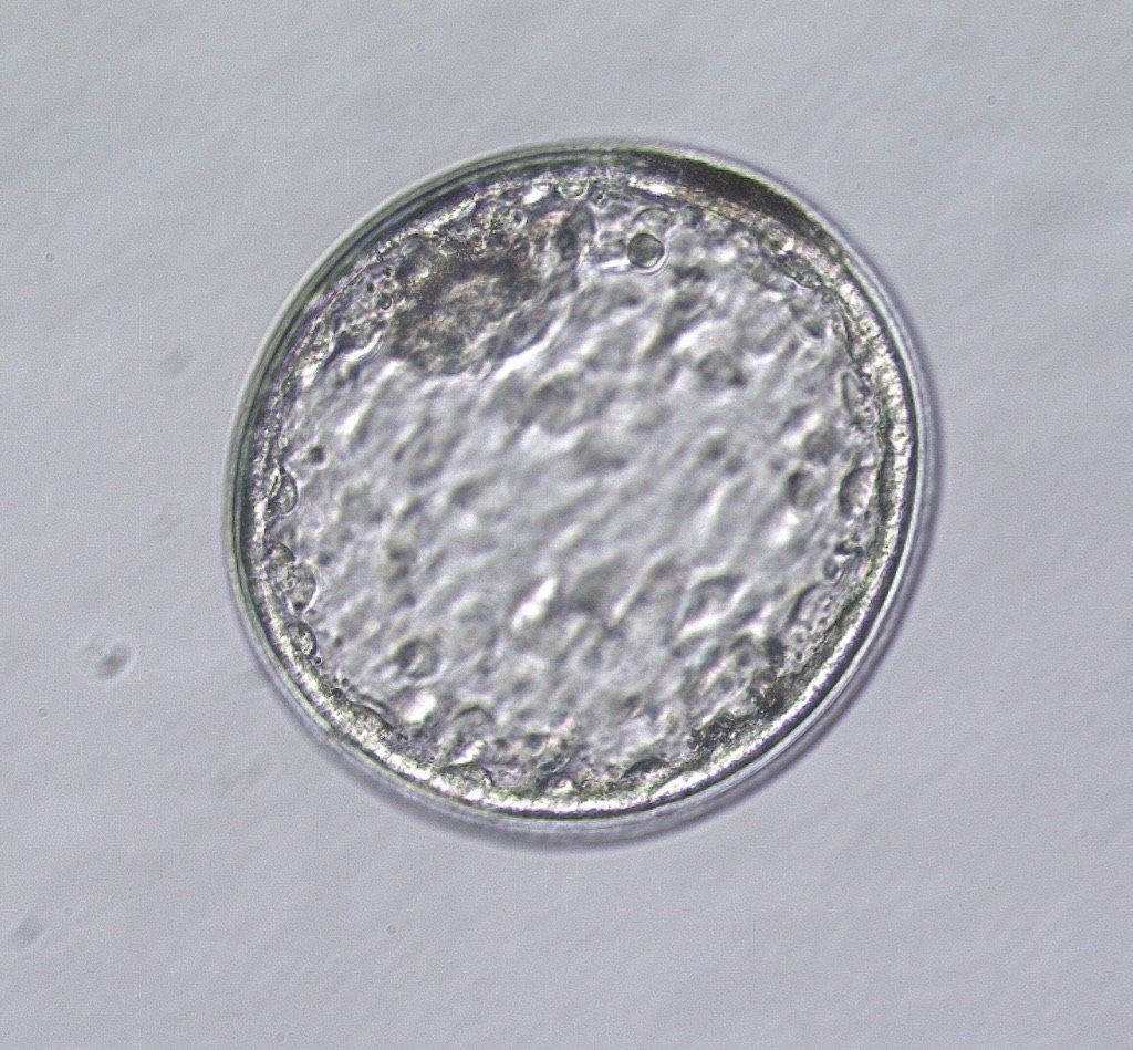
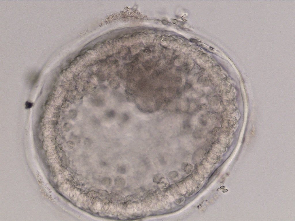
Large Expanded Blastocyst
By day eight, the embryo has increased in size to 800 microns or more and is termed a large expanded blastocyst. At day eight, the embryo will still sink in fluid, but by day 10, it will float. The embryo will nearly double in size each day until day 12.
Grade 1 large expanded blastocyst
Embryo Grading
Embryo grading is an attempt to evaluate abnormalities of individual embryos as they relate to the potential of establishing a pregnancy after transfer. They are graded one through four, with Grade 1 being normal and Grade 4 being severely damaged or degenerated.
Abnormalities in the distribution of fluid are common, and leaks involving the capsule are considered more important than those from the blastocoele.
Unfortunately, grading can only evaluate normal versus abnormal morphology and can in no way evaluate the health of the visible cells. An embryo can have a large number of dead cells and still appear structurally normal until growth or deterioration take place.
Cells that have been dead or stressed for some time will appear darker and are often extruded from the embryo. These are generally visible in the early blastocyst or morula, beneath the zona pellucida.
Grade 1
Grade 1 embryos have few, if any, detectable abnormalities and are expected to have the highest potential for a successful pregnancy. Some minor abnormalities, such as a few extruded blastomeres, are acceptable in a Grade 1 embryo.
Grade 1 expanded blastocyst
Grade 2
Grade 2 embryos have mild abnormalities that are not expected to negatively impact the establishment of a pregnancy, such as in an embryo where fluid has leaked from the blastocoele but is still contained within the capsule.
Grade 2 expanded blastocyst
Grade 3
Grade 3 embryos have significant abnormalities that are likely to result in a decreased chance of pregnancy.
Grade 3 expanded blastocyst
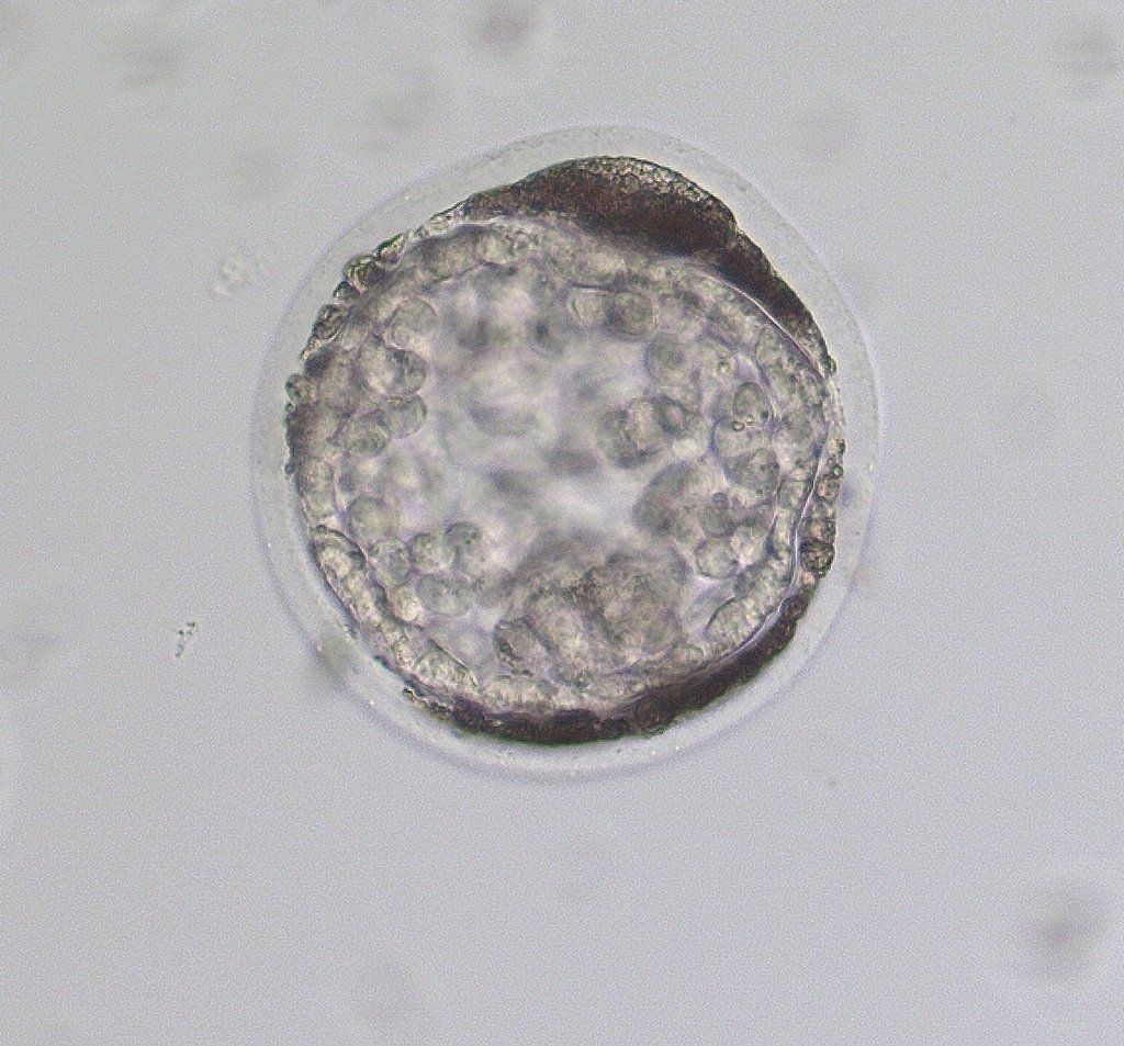
Grade 4
Grade 4 embryos have such significant abnormalities that the establishment of a pregnancy is unlikely.
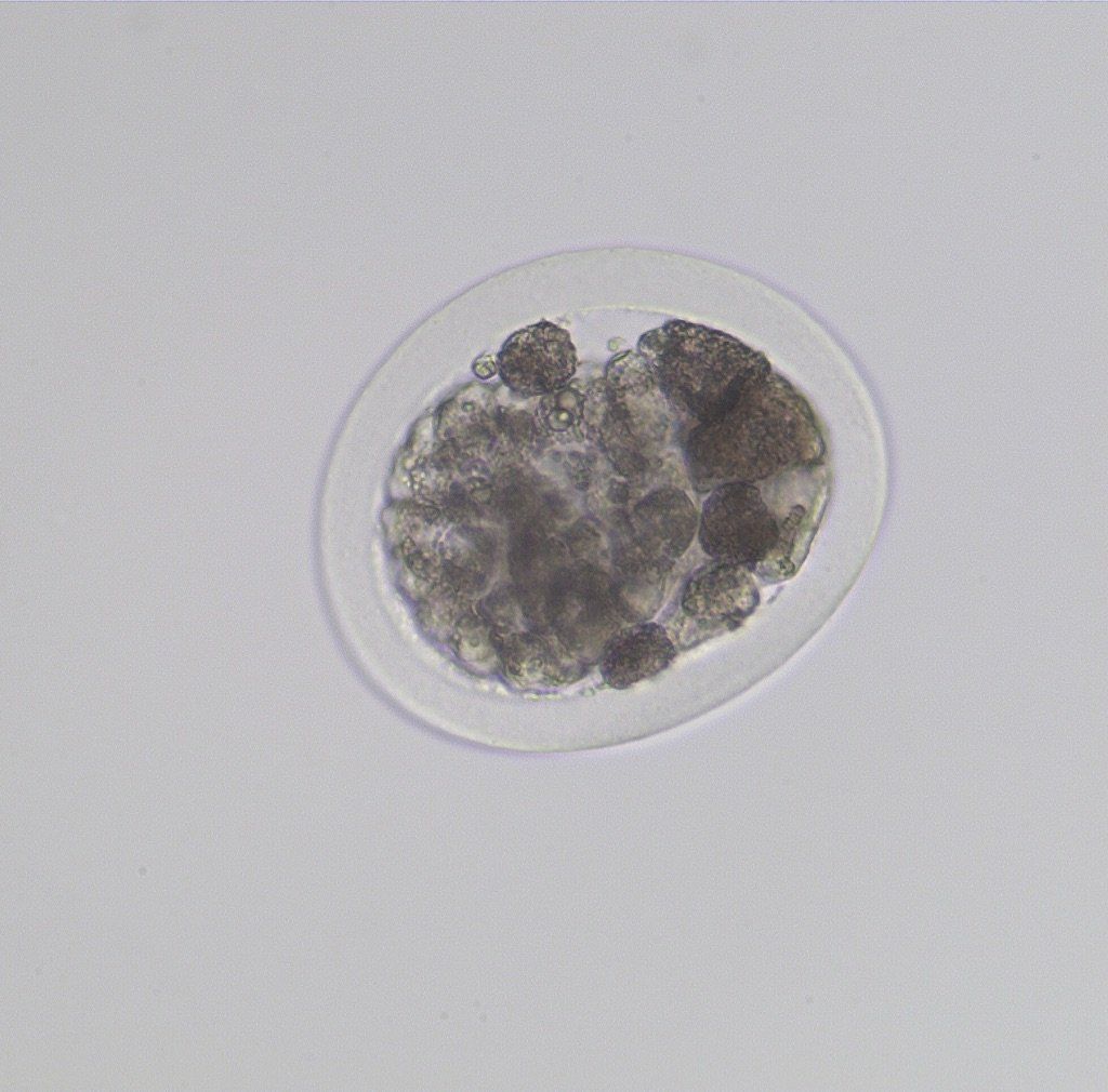
Grade 4 Morula
Reproductive Services
Quick Links
Contract A
In-House ICSI
Contract B
Oocyte Recovery- ICSI Without Recipient
Contract C
Oocyte Recovery, ICSI, ET with Recipient
Contract D
Transported Oocyte - ICSI- Without Recipient
Contract E
Transported Oocyte-ICSI- ET With Recipient
Embryo Transfer